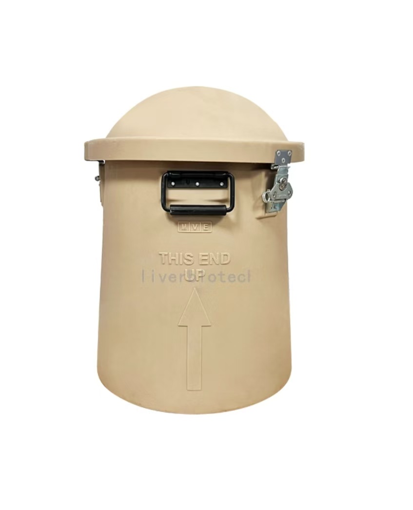
Transport Mode
Liquid Nitrogen Transportation
1、Adsorptive Liquid Nitrogen Transportation ,No free liquid nitrogen ,White vapor emission indicates normal operation.
2、Temperature monitoring devices should closely monitor the temperature inside the tank during transportation. If any abnormalities are detected, you can copy the PDF and Excel data from the temperature recorder. (One end of the temperature monitoring device can be unplugged to reveal a USB connector. Simply insert it into a computer to copy the data. If you encounter any difficulties, you can contact the sales staff and technical support for assistance.)
3、Alloy Combination Lock Designed to ensure no third-party access to the LN₂ tank or cell samples during transportation from dispatch to receipt, thereby safeguarding cell integrity.
4、GPS Tracker Enables real-time tracking of cell transportation routes to prevent loss.
5、Liquid Nitrogen Tank, temperature monitoring device, alloy combination lock, and GPS tracker are returnable components. Please store them properly and avoid damage; failure to comply will result in blacklisting.
Reference Citation
pass over.
Scope of Application
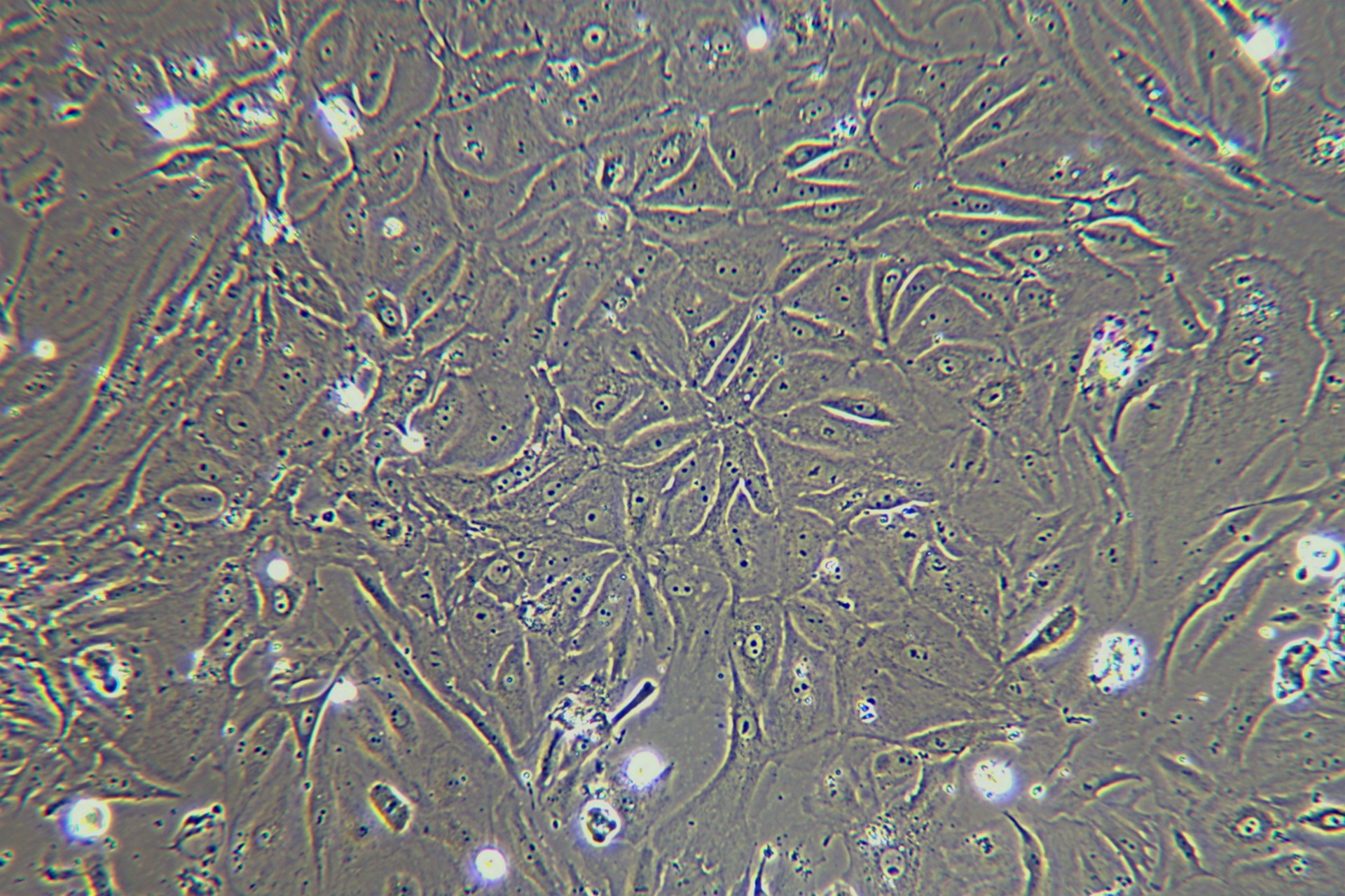
Liver non-parenchymal cells (including hepatic stellate cells, liver sinusoidal endothelial cells, Kupffer cells, biliary epithelial cells, etc.) are critical targets for studying liver function and diseases, and their research applications can be summarized into the following core directions:
1. Mechanistic Elucidation of Liver Diseases
Liver Fibrosis and Cirrhosis: Activated hepatic stellate cells (HSCs) secrete collagen, while liver sinusoidal endothelial cells (LSECs) undergo capillarization, jointly driving fibrogenesis. Kupffer cells exacerbate this process by releasing pro-inflammatory cytokines (e.g., TNF-α, IL-6) that further activate HSCs.
Fatty Liver and Metabolic Disorders: Liver sinusoidal endothelial cells (LSECs) modulate lipid metabolism, Kupffer cells drive inflammatory cascades (e.g., TNF-α/IL-1β release), and hepatic stellate cells (HSCs) exhibit dysregulated vitamin A storage, collectively contributing to non-alcoholic fatty liver disease (NAFLD) progression and insulin resistance.
Liver Cancer Microenvironment: Activated hepatic stellate cells (HSCs) deposit extracellular matrix (ECM) components (e.g., collagen I, fibronectin) to facilitate tumor invasion, while liver sinusoidal endothelial cells (LSECs) sustain tumor growth via pro-angiogenic VEGF signaling. Kupffer cells further orchestrate immune evasion through PD-L1 upregulation and T-cell exhaustion.
2. Immune Regulation and Inflammation Research
Kupffer cells: As the resident macrophages of the liver, they clear pathogens, regulate T-cell tolerance (e.g., through PD-L1 expression), and are studied in the context of viral hepatitis and autoimmune liver diseases.
LSECs (Liver sinusoidal endothelial cells)-mediated immune tolerance: LSECs induce T-cell anergy through antigen presentation, thereby maintaining hepatic immune tolerance. Key research targets include immune checkpoints (e.g., CTLA-4).
Inflammation amplification vs. resolution: Non-parenchymal cells (NPCs) collaboratively secrete pro-inflammatory and anti-inflammatory mediators (e.g., IL-10, TGF-β), thereby modulating inflammatory balance. This mechanism is central to exploring inflammatory equilibrium in sepsis and drug-induced liver injury (DILI).
3. Drug Development and Toxicity Assessment
Drug metabolism models: Liver sinusoidal endothelial cells (LSECs) clear nanodrugs via scavenger receptor-mediated pathways.Hepatic stellate cells (HSCs) in their activated state reduce the efficacy of anti-fibrotic drugs.Cholangiocytes serve as in vitro models for drug screening in cholestatic liver diseases.
Mechanistic studies of toxicity: Kupffer cell activation (e.g., alcohol-induced reactive oxygen species [ROS] release) Oxidative stress responses in hepatic stellate cells (HSCs) These models are used to investigate mechanisms of liver injury caused by environmental toxins or chemotherapeutic agents.
Targeted delivery systems: Designing liver-targeted carriers by leveraging the phagocytic properties of Kupffer cells.
Enhancing drug efficacy through surface-modified nanoparticles that evade scavenger receptor-mediated clearance by liver sinusoidal endothelial cells (LSECs).
4. Regenerative Medicine and Tissue Engineering
Liver regeneration models: Hepatic stellate cells (HSCs) secrete hepatocyte growth factor (HGF) to drive hepatocyte proliferation.
Liver sinusoidal endothelial cells (LSECs) support the regenerative microenvironment through angiogenesis.
Cholangiocytes participate in the remodeling of the biliary network.
Organoids and Organ-on-a-Chip: Bioengineered liver models constructed by co-culturing non-parenchymal cells (e.g., HSCs, hepatocytes, and LSECs) to recapitulate pathological microenvironments such as fibrosis or tumor niches.
Regulation of Stem Cell Differentiation: Non-parenchymal cells (NPCs) secrete signaling factors (e.g., Wnt/β-catenin and Notch pathways) to orchestrate the lineage-specific differentiation of hepatic progenitor cells (HPCs), a critical strategy for advancing cell transplantation therapies in liver regeneration.
5. Inter-organ Interactions and Systems Biology
Gut-liver axis research: Kupffer cells clear gut-derived bacterial metabolites (e.g., lipopolysaccharide, LPS).
Liver sinusoidal endothelial cells (LSECs) orchestrate immune cell trafficking.
These mechanisms elucidate how the gut microbiota influences liver diseases.
Metabolic network regulation: Non-parenchymal cells (NPCs) modulate systemic glucose and lipid metabolism via secreting factors such as FGF21 (fibroblast growth factor 21) and adiponectin, thereby mechanistically linking the pathogenesis of obesity, diabetes, and liver diseases.
Application of Single-Cell Omics: Single-cell RNA sequencing (scRNA-seq) resolves the heterogeneity of non-parenchymal cells (NPCs) (e.g., M1/M2 polarization states in Kupffer cells), uncovering disease-specific cell state transitions.
Conclusion
Research applications of liver non-parenchymal cells (NPCs) span multiple dimensions, including disease mechanisms, immune regulation, drug discovery, regenerative medicine, and inter-organ crosstalk. The study of their functional networks provides critical therapeutic targets and experimental platforms for precision medicine in liver diseases and biotechnological innovations.