|
Type
|
Standard
|
|
Product
|
ECM-Gel
|
ECM-Gel phenol red-free
|
|
Catalog
|
LV-6828-2-05ml
|
LV-6828-2-10ml
|
|
Specification
|
5mL
|
10 mL
|
|
Concentration of protein
|
8-10mg/mL
|
8-10mg/mL
|
|
Free shipping or not
|
Over 3000 free shipping
|
Over 3000 free shipping
|
|
Delivery
|
1 week
|
1 week
|
|
Carriage
|
Transportation of Dry ice
|
Transportation of Dry ice
|
|
Storage
|
-70 ℃ to -20 ℃
|
-70 ℃ to -20 ℃
|
|
Life cycle
|
24 months
|
24 months
|
|
Warranty
|
21 months
|
21 months
|
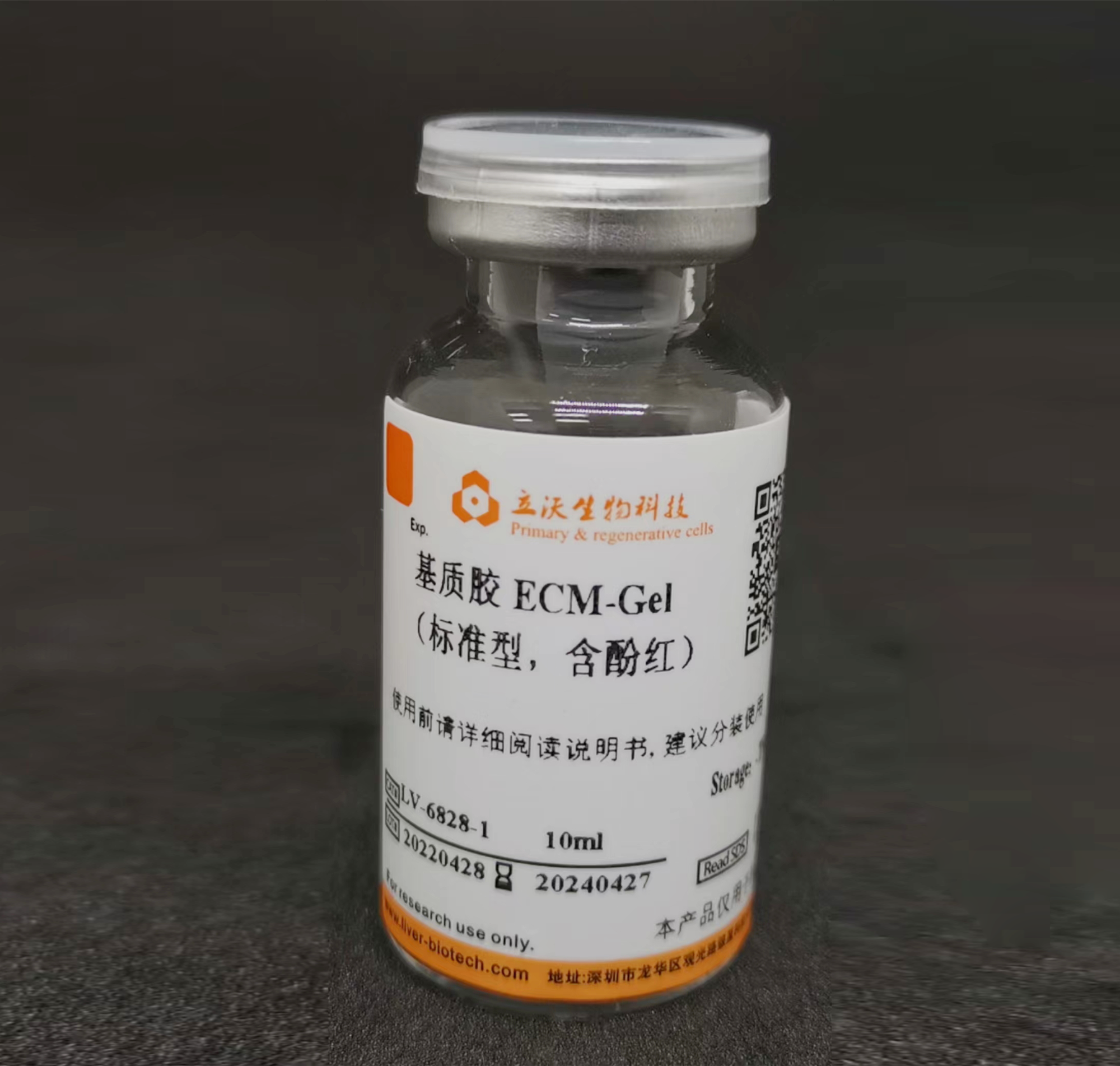
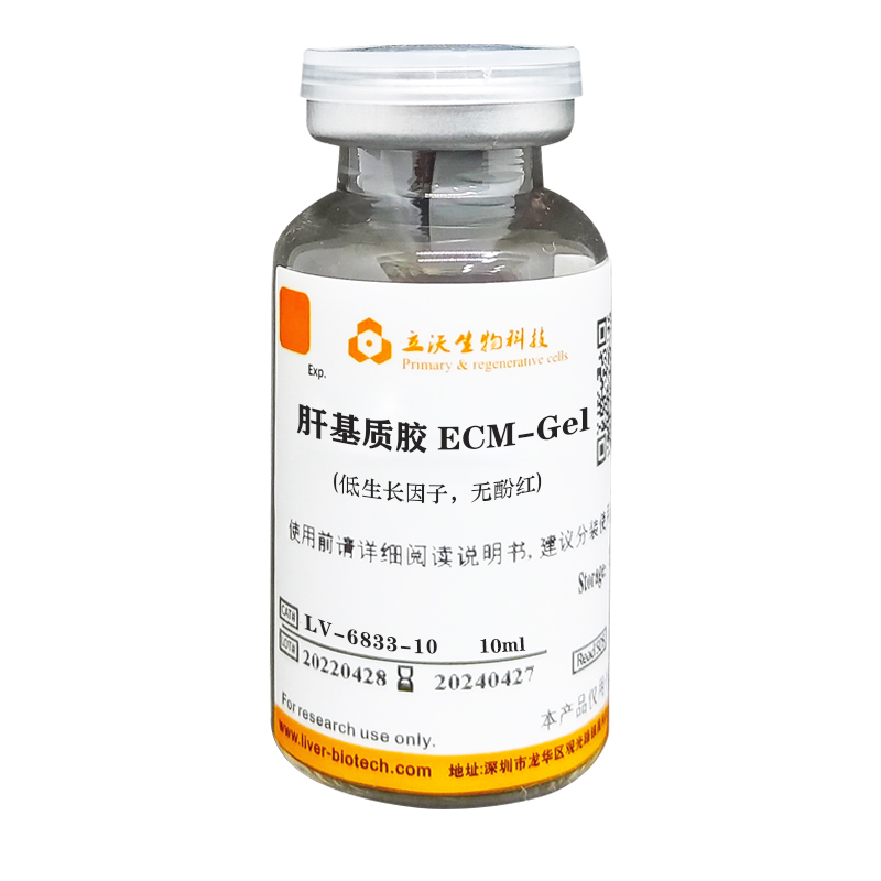
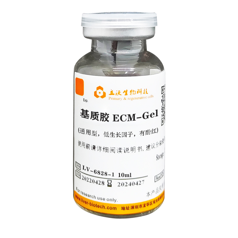
ECM-Gel
Store at -20℃. Avoid repeated freezing and thawing. For Research Use Only.
I Introduction
ECM-Gel is a soluble substrate membrane preparation extracted from healthy pigs, which shapes into colloidal forming above 10℃. Compared to rodents, pigs are closer to humans in terms of genetic distance. ECM-Gel is naturally substitutable and has a wide-spread applications in clinical transplantation, medicine, cosmetic medicine and pharmaceutical auxiliary materials. Moreover, there are hundreds of literatures have been published on Pubmed at present, indicating that the ECM-Gel of pig origin are naturally replaceable. Based on our decellularization technology, as well as standardized, large-scale production and manufacture experience, we ensure the excellent quality, uniformity, consistency and stability of products, offering the scientists another choice.
The major components of this product are laminin, type IV collagen, nestin and heparan sulfate proteoglycan, with the advantages of low endotoxin and DNA content. It is mainly used to support cell growth and differentiation in vitro, metabolic/toxicology studies, cell adhesion, angiogenesis, cells migration/invasion and in vivo tumorigenesis experiments, etc.
ECM-Gel Products and their Applications:
|
Type
|
ECM-Gel
|
Cat.no.
|
Size
|
Applications
|
|
Standard
|
ECM-Gel
|
LV-6828-1
|
5/10ml
|
Cell growth, differentiation, invasion, morphology, function, gene expression, etc. Protein concentration is 8-10mg/ml.
|
|
ECM-Gel
phenol red-free
|
LV-6828-2
|
5/10ml
|
|
Growth Factor Reduced (GFR)
|
ECM-Gel
|
LV-6829-1
|
5/10ml
|
Suitable for higher requirements of ECM-Gel experiments, reduce the interference of cytokines;Protein concentration is 8-10mg/ml.
|
|
ECM-Gel
phenol red-free
|
LV-6829-2
|
5/10ml
|
|
Standard, High Concentration
|
ECM-Gel
|
LV-6830-1
|
5/10ml
|
In vivo experiments, angiogenesis, culture of tumor organoids, etc. Better support for 3D cell culture, cells grow better in 3D environment;Protein concentration is 16-20mg/ml.
|
|
ECM-Gel
phenol red-free
|
LV-6830-2
|
5/10ml
|
|
GFR, High Concentration
|
ECM-Gel
|
LV-6830-3
|
5/10ml
|
|
ECM-Gel
phenol red-free
|
LV-6830-4
|
5/10ml
|
Remarks: Our ECM-Gel products are derived from healthy pigs, not mouse EHS tumor tissue. The matrix collagen composition and proportion are slightly different, and the optimal gelatinization concentration is slightly lower than the Matrix-gel concentration.
II Reagents and Materials
-Serum-free medium
-Vortex mixer
-Pre-chilled centrifugal tube
-Culture plate
-Pipette
-Pre-chilled tip
-Pipettor
-Biosafety cabinet
-CO2 incubator (37℃/5%)
III Methods
Precautions:
1) Adequate melting of the ECM-Gel requires at least 4℃ for overnight. In some cases, the product concentration is high, it takes longer to melt.
2) Throughout the operation, all culture vessels, consumables and mediums in contact with the ECM-Gel should be pre-cooled, as the ECM-Gel will begin to gel at more than 10℃.
3) The optimal concentration of matrix gelatinization in vitro is 6 mg/ml, and the in vivo gelatinization should not be less than 3 mg/ml.
4) For your safety and health, please wear a lab coat, a disposable mask and gloves, and take necessary precautions.
Thawing
Transfer the ECM-Gel product from the -20 °C refrigerator to the 4 °C refrigerator (on ice) for overnight thawing (it may take more time when the protein concentration is high), and shake the vial with vortex after liquefaction to ensure that the ECM-Gel is evenly dispersed. If you can't use it all at a time, you can divide it on ice once and freeze-thaw it only once. If the ECM-Gel has more bubbles, they can be removed by centrifugation at a low temperature of 400g for 5 minutes. Place the ECM-Gel in a sterile area and set it aside on ice.
Caution: Be careful not to over-dry the ECM-Gel during the gelling stage. The melted ECM-Gel will gradually solidify above 10 °C. And the ECM-Gel should always be placed on ice throughout the experimental process.
Gelling
1. Thin layer gel method (thickness 0.5 mm)
1)Use pre-cooled tip or pipette and gently aspirate the ECM-Gel to mix evenly (whenever the ECM-Gel blocks the tip or pipette, please change the tip or pipette). If the ECM-Gel has more bubbles, they can be removed by centrifugation at a low temperature of 400g for 5 minutes.
2)Place the culture plate on ice, and add ECM-Gel to the culture plate according to 50μl of ECM-Gel/cm2 to ensure that the colloid is evenly coated at the bottom of the culture well.
3)Place the culture plate in a 37℃ incubator for 30-40 minutes to make the ECM-Gel completely gelatinous.
4)Gently wash the culture wells with serum-free medium to remove the unsolidified matrix proteins. Make sure that the tip of the pipette does not scratch the glue. The plates are now ready to use.
2. Thin layer coating method (standard/growth factor reduced 8-10mg/ml)
1)Use pre-cooled tip or pipette and gently aspirate the ECM-Gel to mix evenly (whenever the ECM-Gel blocks the tip or pipette, please change the tip or pipette). If the ECM-Gel has more bubbles, they can be removed by centrifugation at a low temperature of 400g for 5 minutes.
2)Transwell coating:
Dilute the ECM-Gel with pre-chilled serum-free medium in the ratio of 1:6-1:8 (protein coating amount of 100-400μg/cm2). Then, add the diluted ECM-Gel to the Transwell chamber, taking the 24-well plate as an example, 50-100 μl can be added to each chamber.. After that, place it in a 37℃ incubator for 30-40 minutes to make the protein to be fully coated.
3)The coated Transwell or culture plate should be hydrated with serum-free culture in a 37℃ incubator for 30 minutes before use , followed by subsequent migration invasion experiments.
4)Digest treated cells (can be starved for 12-24 h first), make a single cell suspension using serum-free medium containing 0.05%-0.2% BSA, and seed into a transwell chamber in the appropriate number of cells per well.
5)Add 600 μL of 10% FBS medium to the lower chamber of the 24-well plate, and avoid the formation of bubbles between the lower culture medium and the small chamber when adding, and the production of air bubbles is easy to weaken or even disappear the chemotactic effect of the lower culture medium.
6)Cultured cells: 12-48h in normal culture (time depends mainly on the ability of cancer cells to invade).
7)Remove the transwell chamber, discard the culture medium in the wells, wash the PBS 2 times, fix the 4% paraformaldehyde for 30 min, and air dry the chamber appropriately; 0.1% crystal violet staining for 15 min, PBS rinsed, gently wiped off the upper layer of unregranted cells with a cotton swab, and randomly selected 5 field statistics under the microscope.
8):The protein coating amount is from 50μg/cm2 to 500μg/cm2 (cm2 is the culture base area). The optimal amount of coating needs to be optimized according to the experience of the researcher or the individual needs of the researcher.
3. Thick layer gel method (1.0 mm, mainly used for 3D cell, organoid culture)
1)Use pre-cooled tip or pipette and gently aspirate the ECM-Gel to mix evenly (whenever the ECM-Gel blocks the tip or pipette, please change the tip or pipette). If the ECM-Gel has more bubbles, they can be removed by centrifugation at a low temperature of 400g for 5 minutes.
2)Place the culture plate on ice and use a pre-chilled tip to add ECM-Gel to the pre-chilled cell suspension (ECM-Gel: the cell suspension volume is 4:1-2:1, the optimal volume ratio is 3:1). Then, gently mix well.
3)Add cell-containing ECM-Gel to the culture plate at 150-200μl/cm2 (cm2 is the culture floor area). Place the plate in a 37℃ incubator for 30-40 minutes to gel.
4)After gelatinization, add fresh medium and incubate in a 37℃ incubator.
Recommendation: Add medium to wash the uncoagulated matrix protein and incubate in a 37℃ incubator for 10 minutes. Then switch to fresh medium to achieve excellent results.
4. Arch formation method (mainly used for 3D cell and organoid culture)
1)Use pre-cooled tip or pipette and gently aspirate the ECM-Gel to mix evenly (whenever the ECM-Gel blocks the tip or pipette, please change the tip or pipette). If the ECM-Gel has more bubbles, they can be removed by centrifugation at a low temperature of 400g for 5 minutes.
2)Add ECM-Gel to pre-chilled cells/tissues, organoid suspensions (ECM-Gel: the cell volume ratio of suspension is 4:1-2:1, the optimal volume ratio is 3:1). Mix gently.
3)Aspirate the matrix glue suspension with the pre-cooled tip and gently add it into the culture well. In order to improve the arching effect, the culture plate can be preheated at 37℃ for 10 minutes.
4)Place at room temperature for 1-2 minutes, quickly upside down the culture plate (the strength needs to be mastered by oneself and the standard is to form a teardrop-like arch). Then, place it in a 37℃ incubator for 30-40 minutes ( Normally, this product can be gelled for 10 minutes, but 5% CO2 may extend the gelling time).
5)After gelled, organoid medium will be added to continue culture.
5. PDX modelling (high concentration of ECM-gel is recommended)
1)Due to the relatively conservative protein species, the ECM-Gel of pig origin has low immunogenicity. In addition, PDX mice are generally immunodeficient. Therefore, the species differences in ECM-Gel have little effect on the establishment of PDX models.
2)Use pre-cooled tip or pipette and gently aspirate the ECM-Gel to mix evenly (whenever the ECM-Gel blocks the tip or pipette, please change the tip or pipette). If the ECM-Gel has more bubbles in the pre-gel solution, they can be removed by centrifugation at a low temperature of 400g for 5 minutes.
3)Add ECM-Gel to pre-chilled cells/tissues, organoid suspensions and mix gently. This product can be rapidly gelatinized in mice. The minimum tested gel-forming concentration is 3mg/ml, and the gelling time is about 10 minutes. The higher concentration of the ECM-gel, the shorter gelling time and the stronger stiffness achieve.
4)Use the pre-chilled tip or needle to aspirate the ECM-cell suspension. Then quickly transplant it into subcutaneous and other sites of mice under anesthesia to avoid colloidal outflow. The injection process can be practiced, and it confirms whether the ECM-Gel has been gelatinized and the gelatinized time in the process.
IV The digestive passage/cryopreservation
When organoids need to be passed on, organoids in the ECM-Gel can be recovered using the organoid recovery solution (LV-ORS001/2) produced by our company.
1. Thawed digestive solution
Thawed digestive solution could be temporarily stored at 4℃ for 1 week or refrozen back to -80℃ or -20℃ (freeze-thaw no more than 3 times). It is recommended to dispense according to the usage of digestive solution when opening a new one to avoid repeated freeze-thawing. Repeated freezing and thawing or long-term storage at 4℃ will reduce the effect of the digestive solution.
2. Digestive process
1) Remove the medium, gently wash with PBS for 1-2 times. Add 300-500μl 37℃-preheated organoid recovery solution to 50μl ECM-Gel.
2) Then, draw the appropriate amount of recovery solution and blow it down from the top of the organoid. Destroy the ECM-Gel in the shape of“米” with a tip, so that the ECM-Gel will be suspended in the recovery solution.
3) Transfer the plate to 37℃ incubator for 20-30 minutes, during which the plate can be gently shake to speed up the digestion process.
4) Determine the digestion degree under a microscope. Excessive digestion may break down organoids into single cells.
Note: If the gel disappears completely, there may be over digested. The digest time should be carefully tested, e.g. the excessive digestion of intestinal organoids may affect the occurrence and function of organoids.
5) Terminate the digestion by adding the culture medium and transfer the organoid suspension to the centrifugal tube.
<