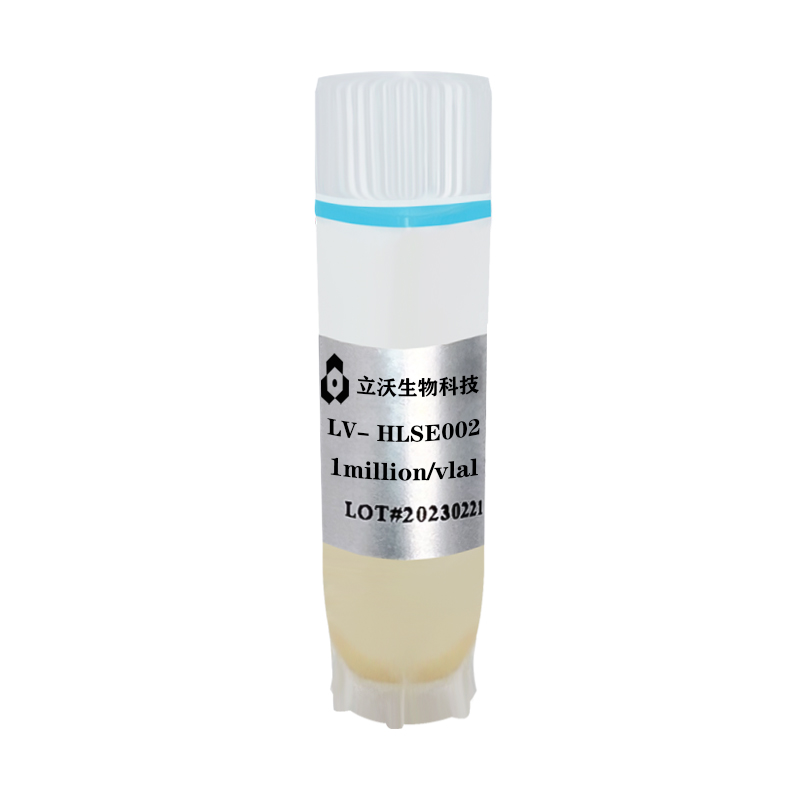
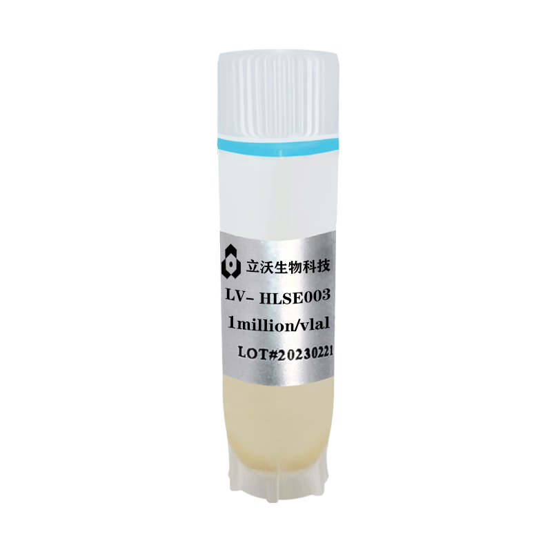
Mouse sinusoidal endothelial cell
(Can be attachment,Cat# LV-MLSE001)
(For research use only)
The instruction is suitable for the mouse sinusoidal endothelial cell of our company. Please follow the instructions,when you use it. If you have any questions, please consult the technicians.
I Introduction
Liver sinusoidal endothelial cells (LSEC) plays an important role in the occurrence of liver physiological functions and pathological mechanisms, and has become a hot spot in liver research in recent years. It was found that LSEC constitutes a semi-permeable barrier between liver parenchymal cells and blood, and is involved in the secretion and regulation of liver metabolism, immune response, growth factors and cytokines in the liver, and plays an important role in the physiological function and pathological mechanism of the liver. This product is isolated into SPF-grade mice by enzymatic perfusion digestion, preserving the biological activity of cells in vivo. Other liver cells are removed by gradient centrifugation with ultra-high purity (>90%). After detection, there is no pollution such as bacteria, fungi, actinomycetes, etc., and the cell viability is high. Our company can personalize the primary cells and isolate different strains or different cells of drug treatment to meet the needs of different customers.
II Reagents and Materials
- Mouse sinusoidal endothelial cell(Cat# LV-MLSE001)
- Recovery medium(Cat#LV-Rec001)
- LSEC culture medium(Cat# LV-MLSEM001)
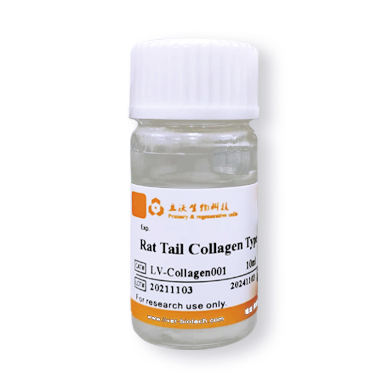
- Collagen-coated culture plate(LV-coated)
- Crushed ice and ice boxes
- Sterile centrifuge tube of 15 ml (Prechilled on ice)
- Disposable pipette
- Wide-mouth pipet tip (The tip of a normal one is cut off and sterilized)
- Pipette
- Thermostat water bath (Preheat at 38℃)
- Refrigerated horizontal centrifuge (with horizontal rotor. it is possible to centrifuge 15 ml of centrifuge tubes)
- Biosafety cabinet
- 37 °C/5% CO2 incubator
- 75% alcohol
III Resurgence and Plating of Cells
1. Insert the 15 ml centrifuge tube into an ice box containing enough crushed ice and sterilize it UV for 15 min. Precool centrifuge.
2. The recovery medium should be placed in crushed ice for sufficiently precooling, and the plating medium should be placed in a 38 °C thermostat water bath to be fully preheated.
3. Quickly transfer the frozen cells from the refrigerated position to a thermostat water bath of 38℃. Then,immerse them in as much water as possible at 38°C and shake clockwise. Please make sure that the cap of the freezing tube is kept above the water.
4. Thaw the freezing tube for about 90-120s, until only a small amount of crushed ice floats in it.
5. Sterilize the freezing tube with 75% alcohol and transfer it to a biosafety cabinet.
6. Resuspend the cells with a wide-mouth pipet tip (gently blow 2 times) and transfer to a precooled centrifuge tube of 15 ml.(Note: if there are more cells left on the freezing tube and the pipette tip, aspirate 1 ml of resuscitation medium to clean the them.
7. Add prechilled recovery medium to the cell suspension dropwise and add 10 ml of recovery medium to every 1 ml of cell suspension (Note: the first 3 ml should be added dropwise and shaken slightly, and the next 7 ml can be accelerated). Finally, slightly upside down 1 time to mix well.
8. Centrifuge of 300 × g at 4 °C for 5 min, remove the supernatant and resuspend with plating medium.
9. The pelleted cells should be resuspended with LSEC culture medium and set the volume to 4 ml. The viability and total amount of cells can be measured by trypan blue exclusion.
10. Inoculate the cells into collagen-coated plate according to the number of 3×10⁴cells/cm 2. Shake the plate well and incubate in a CO2 incubator of 37℃/5%. Then change the solution after 24h
IV Cell Culture and Passage
1. The cells can be passaged when the confluency reaches 80%.
2. Put the medium, PBS and pancreatin into a 37 °C thermostat water bath to preheat, wipe with 75% alcohol and then put it into the ultra-clean table.
3. Aspirate the old culture solution and add a small amount of PBS to wash the cells. In addition, add an appropriate amount of pancreatin so that the amount of pancreatic enzyme can cover the cells. Incubate the cells at 37°C for 2-3 min and observe them under the microscope.
4. Discard the pancreatin when the inter-cell gap becomes larger and the cells tend to be rounded but have not yet floated, and add fresh LSEC culture medium. Then shake the cell flask to terminate the effect of pancreatin, carefully blow the adherent cells with a pipette to make a cell suspension (control the force of blowing to avoid generating a large number of bubbles).
5. Centrifuge of 300 × g at room temperature for 5 min, remove the supernatant and resuspend with medium.
6. Inoculate the cell suspension at the density of 3×10⁴cells/cm2 into a new collagen-coated flask/plate and place it in a 37°C/5% CO2 incubator. Then observe the growth of the adherent every other day.
V Customer Service
If you find any quality problems with the product, please collect the original data and contact the company's salesmen or technical support at the first time. The company ensure after-sales service. Every laboratory has different conditions, different operating habits, different proficiency, and objective factors in experimental failure. If the operation is not strictly in accordance with the instructions or exceeds the time limit of after-sales, the company does not do after-sales. Please understand and support us.
Validity period and raw data provided:
Resuscitation problems: Within 24 hours of resuscitation, trypan blue staining or PI staining should be provided.
Pollution problems: Within 96 hours of resuscitation, microscope photographs of differences should be provided.
Purity issues: Within one month, immunofluorescence or flow cytometry results should be provided
VI Contact Number
Tel:0755-28284050
Marketing representative: 18129812531 (Manager Deng)
Technical Support: 19902901483 (Dr. Zhou)